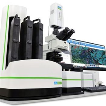
PerkinElmer, Inc., a global leader focused on improving the health and safety of people and the environment, today announced the launch of its Vectra® 3 automated, high-throughput quantitative pathology imaging system. This solution’s new seven color multiplexing and visualization capabilities are designed to enable pathologists and oncologists conducting research to gain a deeper level of understanding of disease mechanisms related to new cancer immunotherapy approaches.
The Vectra 3 system visualizes, analyses, quantifies and phenotypes immune cells in situ in formalin-fixed paraffin-embedded (FFPE) tissue sections, a process that can help provide researchers critical insight into the role of immune cells within solid tumors and the tumor microenvironment. It incorporates 10x whole slide imaging and the Phenochart™ whole slide viewer, allowing researchers to annotate and navigate slides with interactive interfaces to better identify regions of interest for detailed multispectral acquisition.
Unlike alternative instrumentation, limited by the number of colors that can be imaged on one slide, the Vectra 3 system can separate up to seven colors. This multiplexing capability enables identification and quantification of multiple biomarkers and reveals spatial context within a digital workflow to assist researchers with better, faster decisions.
“With its ability to resolve multiple biomarkers in a single tissue section, this new system provides an innovative approach to help researchers more deeply analyze cancer mechanisms and potentially develop breakthrough immunotherapies for better health outcomes,” said Brian Kim, President, Life Sciences & Technology, PerkinElmer. “The information may eventually allow for improved stratification of patients for clinical research, further unlocking the potential of precision medicine.”
The Vectra 3 system is part of PerkinElmer’s Phenoptics™ workflow solutions for quantitative pathology research applications. Phenoptics solutions help researchers fully characterize immune cells and tumor cells in situ within tissue, enabling visualization and analysis of complex cell interactions in ways that are difficult to achieve with other methods.
Combined with PerkinElmer’s inForm® image analysis software, which automatically identifies and quantifies cell populations in tissue, the Vectra 3 system provides researchers the power of multiplexed biomarker imaging and quantitative analysis within a familiar digital workflow. PerkinElmer’s Phenoptics portfolio also includes the Mantra™ quantitative pathology workstation, Opal® research kits, and inForm software.
For more information on PerkinElmer’s Phenoptics solutions, please visit www.perkinelmer.com/cancer-immunology.
