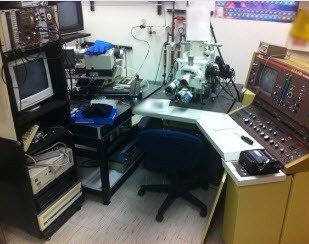
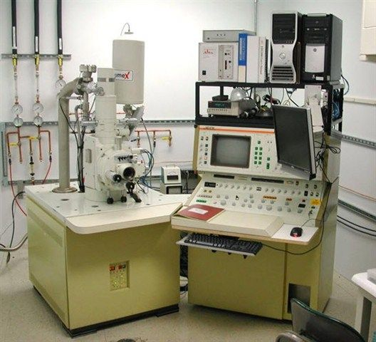
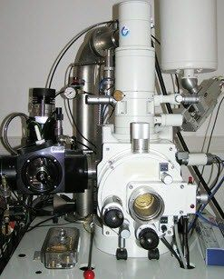
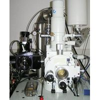
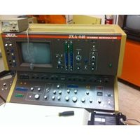
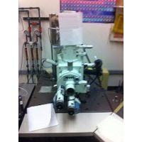
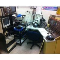
JEOL - JXA-840
Manufactured by JEOL
JSM-840 examines structure by bombarding the specimen with a scanning beam of electrons and then collecting slow moving secondary electrons.
The JSM-840 examines structure by bombarding the specimen with a scanning beam of electrons and then collecting slow moving secondary electrons that the specimen generates. These are collected, amplified, and displayed on a cathode ray tube (CRT, typically a slower version of the picture tube of a television set) although now, most are driven by PCs and these computer-generated images are displayed on LCDs.
The electron beam is scanned using a raster pattern so that an image of the surface of the specimen is formed. Specimen preparation typically includes drying the sample and making it conductive to electricity, if it is not already. Photographs are taken at a very slow rate of scan in order to boost the signal and capture greater resolution. All our SEMs use digital imaging.
SEM is typically used to examine the external structure of objects that are as varied as biological specimens, rocks, metals, ceramics and almost anything that can be observed in a dissecting light microscope.
Active Questions & AnswersAsk a Question
1Replies
JEOL JXA-840
Recent Questions & Answers
Updated byLabWrenchSiteAdmin
Electron Microscopes Service ProvidersView All (8)
Documents & ManualsView All Documents
Features of JXA-840
- 30 Ångstrom resolution (LaB6 source at 40KV)
- Solid state backscattered electron detector
- Kevex X-ray analyzer with light element capabilities and IXRF software and digital imaging capability
- Equipped for x-ray feature analysis, mapping and quantitative analysis
General Specifications
| Magnification | 10 to 300000 x |
| Accelerating voltage | 1000 to 40000 VA |
| Microscope Type | Electron |

