Olympus - FV1000
The Olympus FluoViewTM FV1000 is a next-generation imaging system designed for high-resolution, confocal observation of...
Active Questions & AnswersAsk a Question
Recent Questions & Answers
Asked byNoga
Need Equipment Support?
Documents & ManualsView All Documents
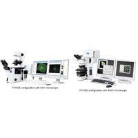
Features of FV1000
Thanks to the variable VBF with 2 nm resolution, acquisition wavelength can be set freely to suit various fluorochromes. Simple separation of fluorescent cross-talk using the lambda scanning and unmixing functions.
Quantification Laser power monitoring for stable stimulation. Stable excitation light via advanced laser intensity feedback system for greater stability. Measurement of fluorescent intensity during observation using live-plot.
High Sensitivity Newly designed Analog Accumulation Circuit (AAC). Uniquely coated filters and dichromatic mirrors enhance sensitivity. Highly sensitive photomultiplier selected specifically for the FV1000. Superior optics with minimum optical return loss.
General Specifications
| Microscope Type | Confocal |
Additional Specifications
Laser Light
Ultraviolet/Visible light laser:
LD laser: 405nm: 50mW, 440nm: 25mW, 473nm: 15mW, 559nm: 15mW, 635mW, 20mW
Multi Ar laser (457nm, 488nm, 515nm, Total 30mW), HeNe(G) laser (543nm, 1mW)
AOTF laser combiner: Visible light laser platform with implemented AOTF system, Ultra-fast intensity modulation with individual laser lines, additional shutter control
Continuously variable (0.1% - 100%, 0.1% increment), REX: Capable of laser intensity adjustment and laser wavelength selection for each region
Fiber: Broadband type (400nm-650nm)
Scanning and Detection
Scanner module: Standard 3 laser ports, VIS - UV - IR
Excitation dichromatic mirror turret, 6 position (High performance DMs and 20/80 half mirror), Dual galvanometer mirror scanner (X, Y)
Motorized optical port for fluorescence illumination and optional module adaptation, Adaptation to microscope fluorescence condenser
Detector module:
Spectral Type Fluorescence Detection:
Standard 3 confocal Channels (3 photomultiplier detectors)
Additional optional output port light path available for optional units
6 position beamsplitter turrets with CH1 and CH2
CH1 and CH2 equipped with independent grating and slit for fast and
flexible spectral detection
Selectable wavelength bandwidth: 1-100nm
Wavelength resolution: 2nm.
Wavelength switching speed: 100nm/msec
CH3 with 6 position barrier filter turret
Filter Type Fluorescence Detector:
Standard 3 confocal Channels (3 photomultiplier detectors)
Additional optional output port light path available for optional units
6 position beamsplitter turrets with CH1 and CH2
CH1 to CH3 each with 6 position barrier filter turret
(High performance filters.)
Filters: High performance sputtered filters, dichromatic mirrors and barrier filters
Scanning Method: 2 galvanometer scanning mirrors
Scanning Modes:
Pixel size: 64 x 64 — 4096 x 4096
Scanning speed: 512 x 512 (1.1 sec., 1.6 sec., 2.7 sec., 3.3 sec., 3.9 sec., 5.9 sec., 11.3 sec., 27.4 sec., 54.0 sec.)
256 x 256 bidirectional scanning (0.064 sec., 0.129 sec.)
0.5 or 1 microsec dwell time per point for fast bidrectional scanning
Spectral Type Fluorescence Detector: X,Y,T,Z,?
Line scanning: Straight line with free orientation, free line, Point scanning
Filter Type Fluorescence Detector: X,Y,T,Z
Line scanning: Straight line with free orientation, free line, Point scanning
Photo Detection Method: 2 detection modes: Analog integration and hybrid photon counting
Pinhole:
Spectral Type Fluorescence Detector: Single motorized pinhole
pinhole diameter ø50 - 300µm (0.5µm step)
Filter Type Fluorescence Detector: Single motorized pinhole
pinhole diameter ø50 - 800µm (0.5µm step)
Field Number (N.A.): 18
Optical Zoom: 1x - 50x in 0.1x increment
Z-drive: Integrated motorized focus module of the microscope, minimum increment 0.01µm or 10 nm
Transmitted Light Detector Unit: Module with integrated external transmitted light photomultiplier detector and 100W Halogen lamp, motorized switching, fiber adaptation to microscope frame
Microscope
Motorized microscope: Inverted IX81, Upright BX61, Upright focusing nosepiece & fixed stage BX61WI
Fluorescence illumination unit: External fluorescence light source with motorized shutter, fiber adaptation to optical port of scan unit
Motorized switching between LSM light path and fluorescence illumination
System Control
PC: PC-AT compatible, OS: Windows XP Professional (English version), Memory: 2GB or larger, CPU:Pentium 3.2GHz or higher, Hard disk: 120GB or larger,
Media: DVD Multi Drive, FV1000 Special I/F board (built-in PC), Graphic board: ATI RADEON X700 PRO
Power Supply Unit:
Spectral Type Fluorescence Detector: Galvo control boards, scanning mirrors and gratings, Real time controller
Filter Type Fluorescence Detector: Galvo control boards, scanning mirrors
Monitor: SXGA 1280X1024, dual 19 inch or larger monitors
Optional Unit
SIM Scanner: 2 galvanometer scanning mirrors, pupil projection lens, built-in laser shutter, 1 laser port
Fiber introduction of near UV diode laser or visible light laser, Optional: 2nd AOTF laser combiner
TIRFM unit: Available laser: 405~633nm. Motorized penetration ratio adjustment. Automatic optical setting for TIRFM objectives.
4th CH detector: Module with photomultiplier detector, barrier filter turret, beamsplitter turret mounted with 3rd CH light path
Fiber port for fluorescence: Output port equipped with FC fiber connector (compatible fiber core 100 -125µm)