HORIBA - XGT-5000
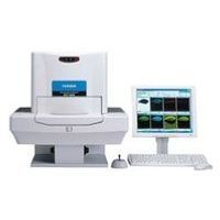
X-ray Analytical Microscope
The sample is visualised with coaxial geometry, so that parallax errors are removed. You have absolute confidence that where you see is where you sample.
Two X-ray guide tubes are provided in the instrument, allowing the user to simply switch between a micro and macro beams, so that a range of experiments can be accommodated. The high intensity beam delivered by these guide tubes ensures that acquisition times are kept to a minimum. The mono-capillary design of these guide tubes are ideally suited for high intensity element imaging, even of samples which are not perfectly flat.
XRF mapped images are easily obtained through automated sample scanning, and the provision of a second detector beneath the sample enables simultaneous acquisition of X-ray transmission images. The additional structural information provided by this technology is extremely useful for locating regions of interest, or interrogating a sample's internal structure.
Active Questions & AnswersAsk a Question
Recent Questions & Answers
Need Equipment Support?
Documents & ManualsView All Documents
Features of XGT-5000
Highest spatial resolution The unique x-ray guide tube technology of HORIBA provides the highest spatial resolution micro-XRF analysis, with x-ray beam diameters down to 10 µm. The high intensity, ultra-narrow beams provided by the guide tubes allow fast, non-destructive analysis of microscopic features.
Transmission X-ray Mapped Imaging In combination with XRF imaging, the XGT-7000 allows transmitted X-ray images to be acquired. This can be used to perform internal structural analyses and identify regions of interest not visible to the eye. Scanning is done with a narrow perpendicular beam, resulting in clear penetrating images even for non-flat samples such as cylindrical parts.
Complete range of sample sizes The accommodating sample chamber enables a wide range of samples to be analyzed, from a 10 µm spot analysis on a microscopic feature, to mapped analysis of areas as large as 10cm x 10cm.
Integrated Data Acquisition and Analysis Software Intuitive software allows easy control of instrument hardware, fast sample visualization and selection of measurement region, and full data analysis. Functions include automated peak identification, quantitative measurements, RGB composite image generation, line profile analysis.
General Specifications
| Depth | 1000 µm |
| Height | 1350 µm |
| Width | 2110 µm |
| Power Requirements | AC100 V/ 50/60 Hz/ 1.3 kVA or less |
| Weight | 280 kg |