Nikon - Eclipse 50i
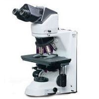
A clinical microscope with the ideal combination of ergonomics, optics and digital imaging capabilities.
Active Questions & AnswersAsk a Question
Recent Questions & Answers
Asked bycal-dave
Need Equipment Support?
Documents & ManualsView All Documents
Features of Eclipse 50i
Rock-solid Stability Utilizing computer-aided engineering (CAE), the new microscopes boast superb durability and stability, even during applications in which they are upgraded with various attachments.
Ergonomic Eyepiece Tube The ergonomic eyepiece tube can be inclined from 10° to 30° and the eyepieces can be extended 40mm. This ensures optimum eye point and a comfortable viewing posture, regardless of the operator's physique or if intermediate modules are used.
Refined Stage with Stay-in-position Stage Handle The refined stage has a stay-in-position stage handle. The stage handle stays at the same position regardless of X-Y stage position. Because the stage handle and focusing knob are always situated close to each other, users can easily and smoothly operate the controls with their hand resting comfortably on the desk. The height and torque of the stage are adjustable to further enhance comfort during operation. Alumite, a new hardening treatment, has been applied to the stage surface to increase durability and smoothness. This facilitates the smooth exchange of specimens while preventing the surface from being scratched by the repeated exchange of glass slides. An additional feature is the refocusing stage. The stage can be lowered by pushing the lever down, and it will return to its original position when the lever is pushed again. This eliminates the need to refocus the image manually each time the specimen is changed and the slide oiled, greatly improving productivity.
Phase Contrast Microscopy Developed expressly for this technique, Nikon's unique Apodized Phase Contrast objectives enable the detection of minute structures which were previously difficult to detect due to annoying halos. Yields excellent contrast and a much wider tonal range and is ideal for urinary sediment tests.
Epi-fluorescence Microscopy A dedicated turret-type epi-fluorescence illuminator has a quick-change mechanism combined with a unique filter-lock system and front-mounted shutter, providing the ultimate combination of performance and convenience in clinical fluorescence diagnostic microscopy.
Ergo-View Cytodiagnosis Unit The Ergo-View cytodiagnosis unit has been developed for easier and more comfortable cytology examinations, allowing fast and accurate motorized magnification changeover with hand switch. A unique quiet, vibration-free mechanism for magnification change ensures superb parfocality of images with no deviation in focusing, and easy slide marking while observing the specimen through eyepieces. Quick exchange of slides with one hand is possible by using an optional specimen holder for one slide.
Easy Access Controls Frequently used controls and switches for adjusting the field diaphragm and illumination intensity have been concentrated in the lower part of the right-hand side to minimize the operator's hand movements and enable operation without having to take your eyes off the specimen.
Renowned Nikon CFI60 Optics Highly acclaimed optics combine the CF design with infinity optics utilizing a 60mm parfocal distance. The results are objectives with longer working distances and high NA's, producing crisp, clear images with minimal flare. The CFI60 optics are perfect for both observations through the eyepiece as well as capturing images with a digital camera. CFI60 optical design provides a flexible upgrade path to accommodate various accessories to be added to the microscope to meet individual applications.
Optional DSC Port An optional DSC port can be combined with an ergonomic tilting/telescoping tube to balance user needs for both digital-image capture and comfortable viewing. Two models are available: a 0.7x lens that is designed to optimize the image to the 2/3-inch CCD, and a 1.0x lens that is designed to optimize the image to a 1-inch CCD. By matching the correct DSC port magnification to the CCD, the same area that is viewed through the eyepieces can be captured with a C-mount digital camera. A centering and focus adjustment mechanism is also provided.
Simple Polarizing/Sensitive Color-Polarizing Microscopy Simple polarizing microscopy can be performed easily using dedicated accessories. Sensitive color-polarizing microscopy enables uric acid crystals forming inside an organism to be identified by changing the interference color via a lever. These components are ideal for gout tests.
Darkfield Darkfield microscopy is ideal for observing specimens such as blood and minute structures like flagella. A dry- or oil-type darkfield condenser can be selected.
General Specifications
| Microscope Type | Upright |
Additional Specifications
Magnification:
10 - 1500X
Optical system:
CFI60 Infinity Optical System
Coarse/fine focusing:Fine: 0.1mm per rotation, Coarse: 13.8mm per rotation, Minimum reading: 1µm; Coarse motion torque adjustable, Refocusing function (with stopper)
Illumination:
6V-30W halogen lamp, 100-240V (worldwide voltage)
Built-in filter:
ND8
Eyepiece tube:
Binocular tube B (for F.O.V. 22mm); Trinocular tube "F" UW (for F.O.V. 22mm/25mm, observation/photo: 100/0, 0/100); Trinocular tube "T" UW (for F.O.V. 22mm/25mm, observation/photo: 100/0, 20/80, 0/100); Ergonomic binocular tube (for F.O.V. 22mm, inclination:10-30°, extension: 40mm); DSC port: 100/0, 50/50 (optional)
Eyepiece lens:
10x (F.O.V.: 22mm), 10x M photo mask (F.O.V.: 25mm), 12.5x (F.O.V.: 16mm), 15x (F.O.V.: 14.5mm), UW 10x (F.O.V.: 25mm), UW 10x M photo mask (F.O.V.: 25mm)
Nosepiece:
Sextuple nosepiece; Ergo-View cytodiagnostic unit (Motorized changeover 10x to 40x with hand switch, Stamp type marking
Stage:
Super-hard Alumite coated surface, Stay-in-position stage handle, Stage handle height and tension adjustable, Rectangular 159 x 243mm surface stage, 78 x 54mm cross travel (x-y movement), 1-slide or 2-slide specimen holder available (option)
Condenser focusing stroke:
27mm
Intermediate accessories:
Epi-fluorescence illuminator (4 filter positions), Magnification module, Eye-level riser, Double port, Teaching head
Observation method:
Brightfield, Epi-fluorescence,Darkfield, Phase contrast, Simple polarizing